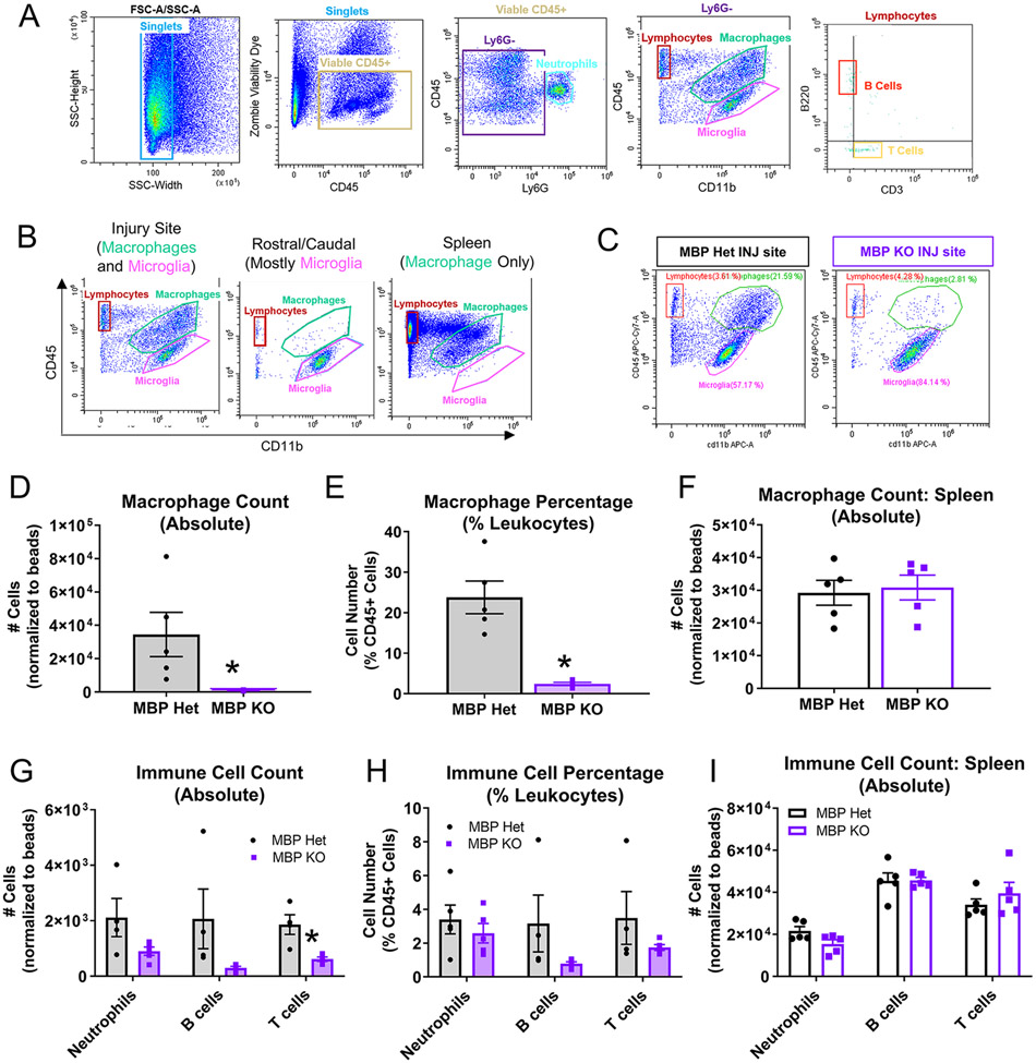
Figure 5. Reduced leukocyte infiltration in MBP KO mice at 7 dpi.
(A) Gating strategy for immune cell flow analysis: viable CD45+ singlets were separated by Ly6G expression to identify neutrophils (Ly6G+/CD45+). Ly6G−CD45+ cells were separated by CD45 and CD11b expression to distinguish macrophage (CD11b+CD45hi) from microglia (CD11b+CD45lo) populations, and to identify CD45+CD11b− lymphocytes. Lymphocytes were separated into B220+ B cells and CD3+ T cells. (B) Spinal cord tissues rostral/caudal to the injury site were used to determine gating for microglia, and spleen was used for gating macrophages. (C) Representative flow plots showing macrophage and microglia populations in the MBP Het versus MBP KO injury site (7 dpi). (D, E) Macrophage number and percentage decreased in MBP KO injury site (n=5 biological replicates per group, *p≤0.05 compared to Het, Two-tailed Student’s t-test, error bars=SEM). (F) Number of splenic macrophages were not different between MBP KO and Het (n=5 biological replicates per group, *p≤0.05 compared to Het, Two-tailed Student’s t-test, error bars=SEM). (G, H) Reduced lymphocyte number and percentage in MBP Het injury site (biological replicates and statistics same as in D, E). (I) Number of splenic lymphocytes were not different between MBP KO and Het (biological replicates and statistics same as in F)

