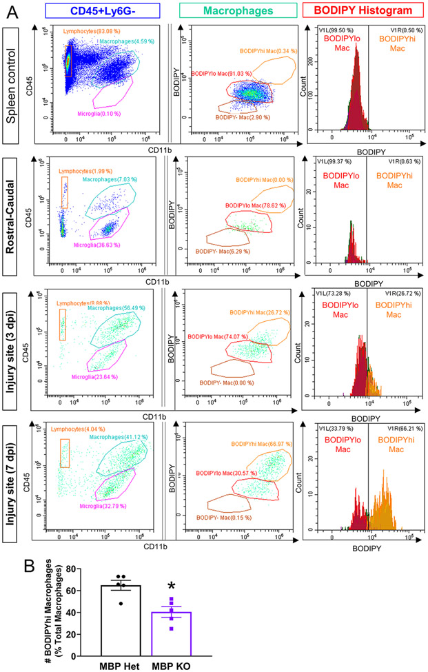
Figure 6. Reduced foamy macrophages in MBP KO injury site at 7 dpi as assessed by flow cytometry.
(A) BODIPY lipid droplet fluorescence in macrophages (CD11b+CD45hi) from spleen, spinal cord tissue rostral-caudal to the injury site, and injury sites from 3 and the 7 dpi. BODIPYhi macrophages are present only after SCI, and increases from 3 to 7dpi. (B) Quantification of BODIPY-hi macrophages at 7 dpi expressed as percent of total macrophages.

