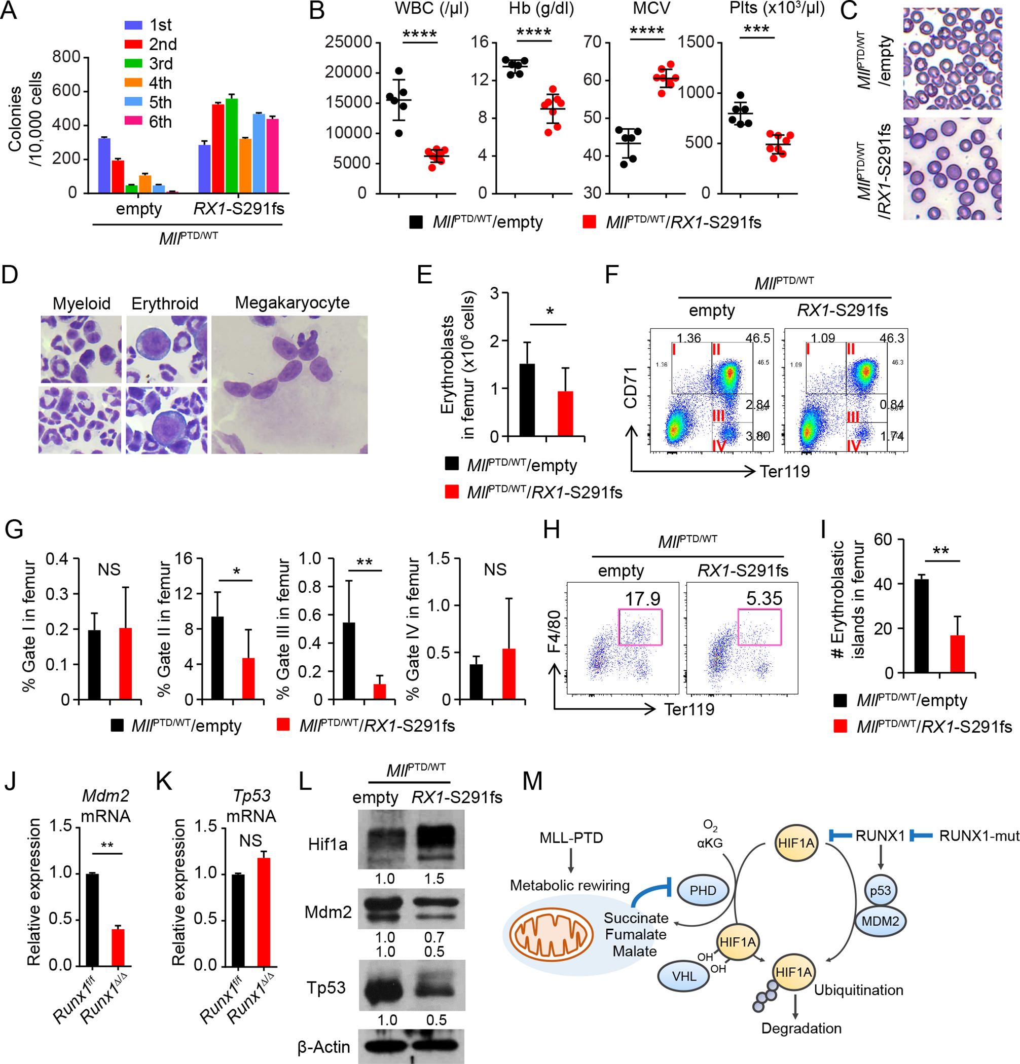
Figure 5. MLL-PTD and RUNX1 Mutations Cooperate to Induce MDS Phenotypes.

A, Serial CFU replating assay of BM cells from indicated genotypes. Data are mean ± s.d. from triplicate cultures. B, WBC counts, HB, MCV, and Plts counts in PB from the MllPTD/WT/empty (n = 6) and MllPTD/WT/Rx1-S291fs mice (n = 8) at 8 weeks after BMT. C, Morphology of RBC in PB smear after fixation. D, Multi-lineage dysplasia in the BM from MllPTD/WT/Rx1-S291fs mice. E, Absolute number of erythroblasts (CD45−CD71high and CD45−Ter119+ cells) in femur from MllPTD/WT/empty (n = 4) and MllPTD/WT/Rx1-S291fs mice (n = 6). F-G, Flow cytometric analysis of 7AAD− CD45− single BM cells from MllPTD/WT/empty (n = 4) and MllPTD/WT/Rx1-S291fs mice (n = 6) (F). Percentages of cells of each fraction in femur are shown in (G). H-I, Flow cytometric analysis of 7AAD− Gr-1− 115− multiplets from MllPTD/WT/empty (n = 3) and MllPTD/WT/Rx1-S291fs mice (n = 3) (H). Absolute number of cells in Ter119+F4/80+ fraction in femur are shown (I). J-K, Mdm2 mRNA (J) and Tp53 mRNA (K) expression in c-Kit+ BM cells from Runx1Δ/Δ mice and control mice. Data are mean ± s.d. from duplicate samples. Results are representative of three independent experiments. L, Hif1a, Mdm2, and Tp53 protein expression in the c-Kit+ cells from indicated mice. M, Model for mechanisms of HIF1A protein stabilization and activation by MLL-PTD and RUNX1 mutant. *P < 0.05, **P < 0.01, ***P < 0.001, ****P < 0.0001; NS, not significant.
