Figure 1.
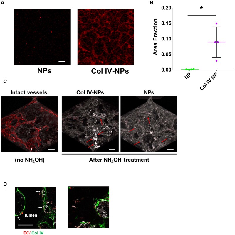
Collagen IV-targeting nanoparticles adhere in vitro to basement membrane produced by endothelial cells in both 2D and 3D settings. (A and B) Compared to non-targeting NPs, Col IV-NPs showed 40-fold increased adhesion to HSVEC-produced basement membrane on a glass slide (n = 4). (scale bar = 100 µm) (C) When tested in a 3D setting, we observed a tubule-shaped binding for Col IV-NPs and a more diffuse deposition for the non-targeting NPs (red arrows). (D) DsRed-labelled ECs (in red), Col IV (in green), and Col IV-NPs (in white) reveals co-localization of NPs at sites of exposed Col IV after EC removal. Mann–Whitney test. *P < 0.05.

