Figure 4.
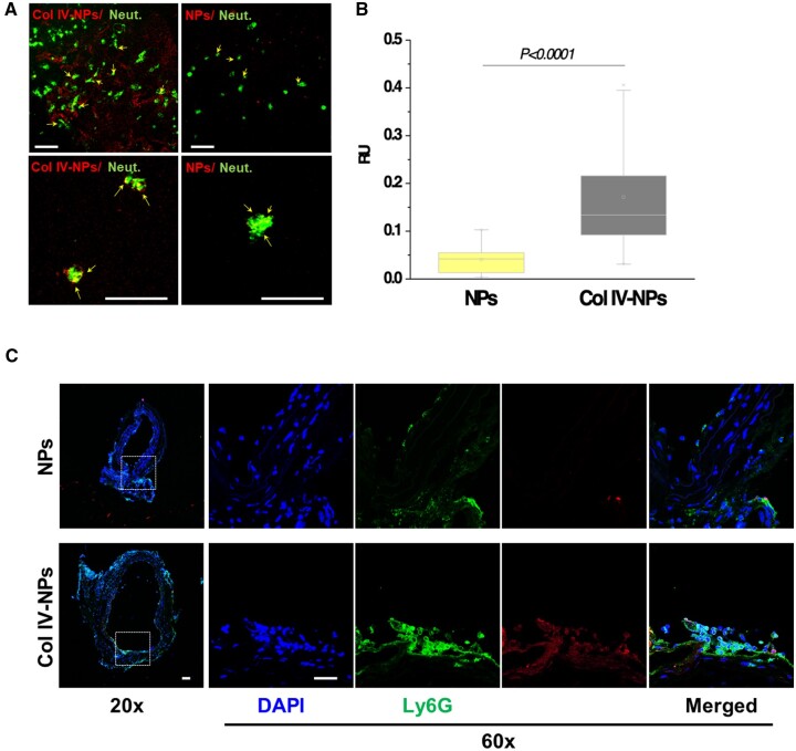
In vitro evaluation of Col IV-NPs targeting to basement membrane. (A) Confocal images of bare and Col IV-NPs (in red) incubated with primary mouse neutrophils (in green) in conditions that mimic superficial erosion. Arrows show NPs internalized in neutrophils (scale bar = 100 µm). (B) Quantitative assessment of NP/neutrophil fluorescent co-localization (n = 24 per group). (C) Immunofluorescent staining of Ly6G (neutrophil marker—in green) shows NPs localization downstream of the flow perturbation. Col IV-NPs co-localize with neutrophils recruited at areas of exposed basement membrane recapitulating eroded plaques. (Scale bar = 25 µm). Mann–Whitney test. P < 0.0001.

