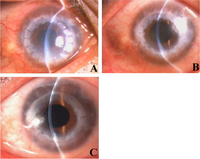
Fig. 1.

A: Representative case #1. Two months after the therapeutic penetrating keratoplasty using cryopreserved tissue, slit-lamp microscopy revealed significant edema and cloudiness in the corneal graft with mild conjunctival hyperemia. B: Representative case #1. Seven months after the therapeutic penetrating keratoplasty using cryopreserved tissue, slit-lamp microscopy showed that the graft had become comparatively clear but still retained a certain degree of edema, making the corneal graft obviously thicker than normal. C: Representative case #1. Ten months after the therapeutic penetrating keratoplasty using cryopreserved tissue, slit-lamp microscopy showed that the graft had become completely clear with normal corneal thickness
