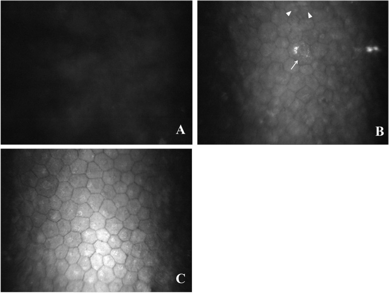
Fig. 3.

A: In vivo confocal microscopy of the corneal graft for representative case #1 at 2 months after therapeutic penetrating keratoplasty. Only a uniformly gray light reflection was observed, and no cells were detected on the posterior surface of the graft. B: In vivo confocal microscopy of the corneal graft for representative case #1 at 7 months after therapeutic penetrating keratoplasty. In vivo confocal microscopy revealed a formed endothelium with multiple nuclei in a single cell (triangle) and cell division morphology (arrow). The cell size and morphology varied markedly. C: In vivo confocal microscopy of the corneal graft for representative case #1 at 10 months after therapeutic penetrating keratoplasty. In vivo confocal microscopy revealed hexagonal and polygonal-shaped endothelial cells on the corneal graft, with a cell density of 1023 cells/mm2. The endothelial cells became relatively consistent in morphology and size
