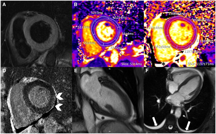
Figure 5.
Anti-PD-L1 antibody-induced myocarditis and pericarditis with moderate to high elevations in high-sensitivity troponin I. (855 ng/L at time of cardiovascular magnetic resonance). (A) T2-weighted SPAIR, (B) T2 mapping, (C) native T1 mapping, (D) phase sensitive inversion recovery late gadolinium enhancement imaging, and (E, F) cine steady state free precession. Very subtle subepicardial late gadolinium enhancement with additional pericardial enhancement (arrowheads), mild pericardial effusion (*), and mild pleural effusions are demonstrated.

