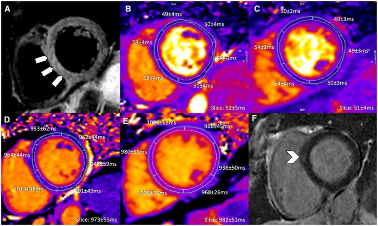
Figure 6.
Anti-PD-L1 therapy-mediated myocarditis with moderate increase in high-sensitivity troponin I (4717 ng/L at time of cardiovascular magnetic resonance). (A) T2-weighted SPAIR, (B, C) T2 mapping, (D, E) native T1 mapping, and (F) phase sensitive inversion recovery late gadolinium enhancement imaging. Diffuse septal oedema (block arrows) and only subtle focal septal late gadolinium enhancement (arrowhead) visualized.

