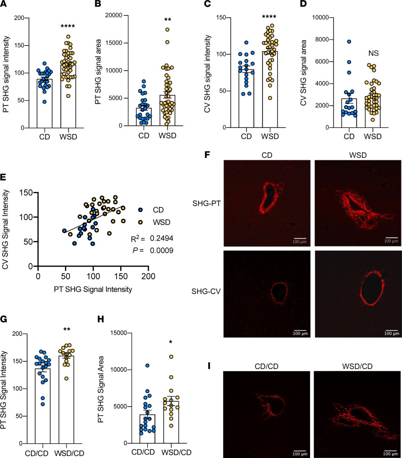
Figure 1. Maternal WSD exposure increases fibrosis in NHP fetal and 1YO offspring liver.
SHG signal intensity (A) and signal area (B) of CD (blue) and WSD (yellow) portal triads (PT) in fetal livers; n = 24 C and n = 40 WSD PTs. SHG signal intensity (C) and signal area (D) of central veins (CV) in fetal livers; n = 18 CD and n = 37 WSD CVs. (E) Correlation between SHG signal intensity of PTs and SHG signal intensity of CVs in fetal livers. (F) Representative SHG images of PTs and CVs in fetal livers, with red indicating SHG signal. Scale bar: 100 μm. SHG signal intensity (G) and signal area (H) of 1YO offspring liver PTs; n = 19 CD/CD and n = 14 WSD/CD PTs. (I) Representative SHG images of PTs in CD/CD and WSD/CD 1YO livers, with red indicating SHG signal. Scale bar: 100 μm. Unpaired 2-tailed Student’s t test was used to test significance. *P < 0.05, **P < 0.005, ****P < 0.0005.

