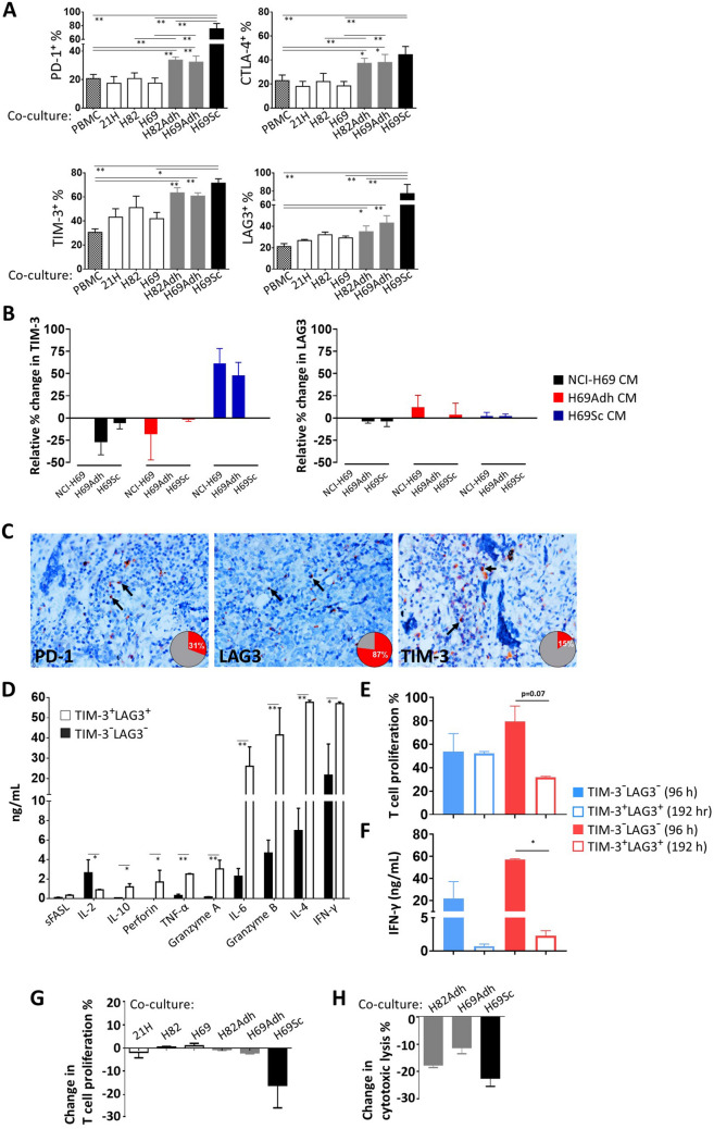
Fig. 4.
Expression of inhibitory receptors and the functional status of T cells co-cultured with SCLC cells. a Expression of PD-1, CTLA-4, TIM-3 and LAG3 on CD8+ T cells in the co-cultures of PBMCs and SCLC subpopulations. The cultures were established in the presence of anti-CD3 mAb (25 ng/mL) for 96 h at a 0.25:1 SCLC:PBMC ratio. b Relative expression change of TIM-3 and LAG3 on CD8+ T cells in the co-cultures of PBMCs and SCLC subpopulations treated with conditioned media collected from different subpopulations in 1:1 ratio. The cultures were established in the presence of anti-CD3 mAb (25 ng/mL) for 96 h at a 0.25:1 SCLC:PBMC ratio with CM collected after 48 h of cell culture. c Representative LAG3, PD-1 and TIM-3 immunohistochemical staining micrographs with checkpoint receptor expressing lymphocytes positive tumor-draining lymph node percentage in SCLC patients (magnification, 400x). TIM-3−LAG3− and TIM-3+LAG3+ CD8+ T cells were purified from the 96 h or 192 h co-culturing of PBMCs and H69Sc cells, d cytokines produced were assessed by a flow cytometric bead array following 16 h stimulation with PMA and ionomycin upon 96 h, e proliferation capacity was assessed by CFSE dilution upon 72 h stimulation with anti-CD3 and anti-CD28 mAbs, f secreted IFN-γ amount with 16 h stimulation with PMA and ionomycin. CFSE-labelled PBMCs were co-cultured with IFN-γ-pretreated or control SCLC cells; the change g in CTL proliferation for 96 h and h CD3/CD28 dynabeads activated CTL co-cultured with IFN-γ-pretreated or control SCLC cells in SCLC cell lysis for 72 h was calculated. (PBMC, control PBMC alone; SCLC-21H, 21H; NCI-H82, H82; NCI-H69, H69; n ≥ 3, *p < 0.05, **p < 0.01)

