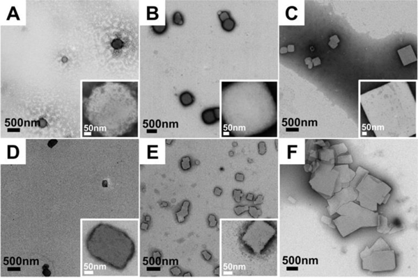
Figure 4.
TEM images of designed sequence assembled at 50°C and observed at different time points. Insets are higher magnification images of individual peptide plates present at each time point. A-C: Peptides assembled at 50°C, pH8 for 15 min, 30 min and 6 hours, respectively. D-F: Peptides assembled at 50°C pH7 for 15 min, 30 min and 6 hours, respectively. Both conditions reveal quick formation of early-time platelet nanostructure. The platelets present at early time points in pH7 eventually fuse together into larger, compound plates. All the images were observed with 2 wt% phosphotungstic acid negative staining to enhance the contrast.

