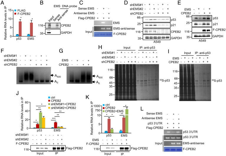
Fig. 5.
EMS coordinates with CPEB2 to inhibit p53 expression. (A) Lysates from A549 cells expressing control or Flag–CPEB2 were immunoprecipitated with anti-Flag antibody. RNAs in immunoprecipitates were then analyzed by real-time RT-PCR to examine EMS and p53 RNA levels. Data shown are mean ± SD (n = 3). The input and immunoprecipitates were also analyzed by western blotting with anti-Flag antibody (SI Appendix, Fig. S3B). *P < 0.05. (B) Lysates from A549 cells were incubated with either sense or antisense biotin-labeled DNA oligomers corresponding to EMS, followed by the pull-down experiments using streptavidin-coated beads. The pulled-down complexes were analyzed by western blotting. The same complexes were also subjected to real-time RT-PCR analysis to examine EMS and p53 RNA levels (SI Appendix, Fig. S3C). (C) In vitro–synthesized EMS or its antisense RNA was incubated with purified recombinant Flag–CPEB2 bound with M2 beads. The inputs and beads-bound RNAs were analyzed by RT-PCR. The primers used for EMS antisense/sense detection do not discern between sense and antisense EMS. (D) A549 cells were infected with lentiviruses expressing EMS shRNA#1, EMS shRNA#2, or CPEB2 shRNA in the indicated combination. Forty-eight hours after infection, cell lysates were analyzed by western blotting. (E) A549 cells were infected with lentiviruses expressing control, EMS, Flag–CPEB2, or EMS plus Flag–CPEB2. Forty-eight hours later, cell lysates were analyzed by western blotting. (F) A549 cells were infected with lentiviruses expressing EMS shRNA#1, EMS shRNA#2, or CPEB2 shRNA in the indicated combination. (G) A549 cells were infected with lentiviruses expressing control, EMS, Flag–CPEB2, or EMS plus Flag–CPEB2. Forty-eight hours after infection, the poly(A) tail length of p53 mRNA was examined using the LM-PAT assay. (H) A549 cells were infected with lentiviruses expressing EMS shRNA#1, EMS shRNA#2, or CPEB2 shRNA in the indicated combination. (I) A549 cells were infected with lentiviruses expressing control, EMS, Flag–CPEB2, or EMS plus Flag–CPEB2. Forty-eight hours after infection, cells were pulse labeled with [35S]cysteine/methionine for 90 min. Cell lysates were then immunoprecipitated with anti-p53 antibody and analyzed by SDS-PAGE, followed by autoradiography. The band intensities of immunoprecipitated p53 in H and I were quantified using ImageJ software and are presented in SI Appendix, Fig. S3 F and I, respectively. (J) A549 cells were infected with lentiviruses expressing control, Flag–CPEB2, Flag–CPEB2 plus EMS shRNA#1, or Flag–CPEB2 plus EMS shRNA#2. Forty-eight hours later, cell lysates were immunoprecipitated with anti-Flag antibody. RNAs present in immunoprecipitates were analyzed by real-time RT-PCR. Data shown are mean ± SD (n = 3). The input and immunoprecipitates were also analyzed by western blotting. *P < 0.05; **P < 0.01; ***P < 0.001. (K) A549 cells were infected with lentiviruses expressing control, Flag–CPEB2, or Flag–CPEB2 plus EMS. Forty-eight hours later, cell lysates were immunoprecipitated with anti-Flag antibody. RNAs present in immunoprecipitates were analyzed by real-time RT-PCR. Data shown are mean ± SD (n = 3). The input and immunoprecipitates were also analyzed by western blotting. *P < 0.05; **P < 0.01. (L) Purified recombinant Flag–CPEB2 bound with M2 beads was incubated with in vitro–synthesized p53 3′-UTR, EMS, and its antisense RNA in the indicated combination. The inputs and beads-bound RNAs were analyzed by RT-PCR. The primers used for EMS antisense/sense detection do not discern between sense and antisense EMS. ctrl, control; IP, immunoprecipitation.

