Fig. 1.
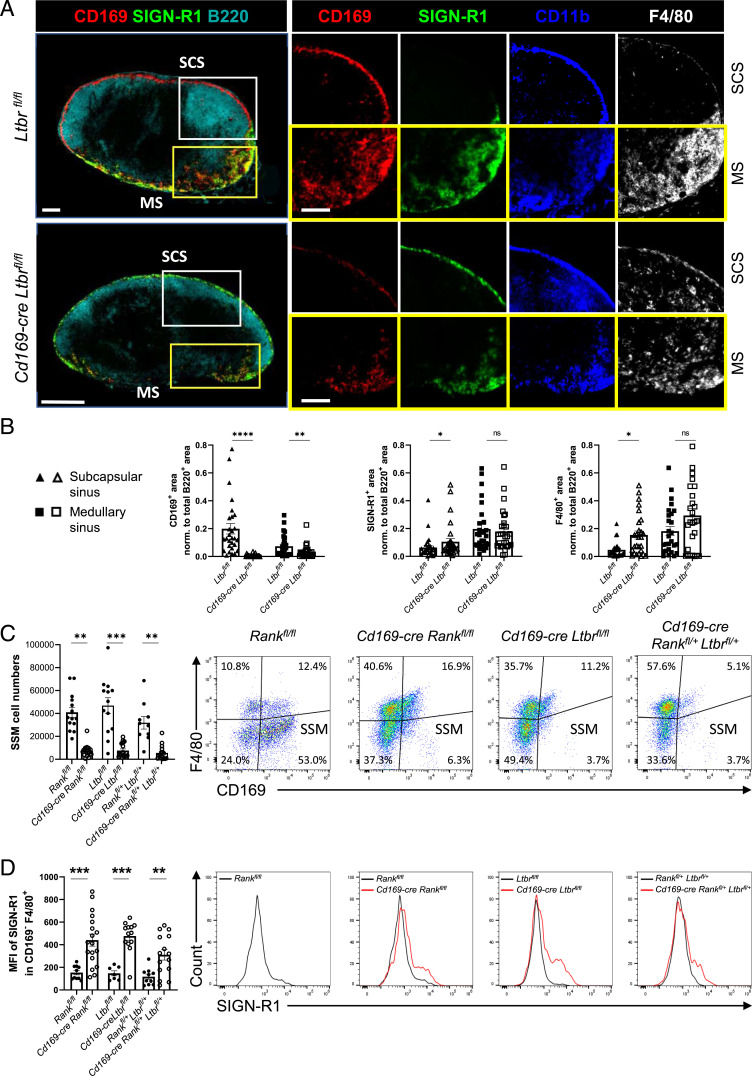
Deficiency in LTβR results in loss of CD169+ SSMs. (A) Wide-field immunofluorescence microscopic images for CD169, SIGN-R1, CD11b, F4/80, and B220 of inguinal LN sections from Ltbrfl/fl and Cd169-cre Ltbrfl/fl mice. White-framed images are higher magnification of the subcapsular sinus (SCS), while yellow-framed images are from the medullary area (MS). Scale bars, 200 µm. (B) Quantification of the area of CD169, SIGN-R1, and F4/80 staining in the SCS and MS normalized to total B220+ area. Shown are the mean and individual data points from auricular and brachial LNs. Statistical significance (Mann-Whitney test); P < 0.005; ns, not significant; error bar, SEM. (C) Flow cytometry analysis of SSMs of inguinal and brachial LNs pregated as live CD11b+ CD11c+ MHC-IIInt cells in mice of the indicated genotype. The proportion of the cells in the quadrants is indicated. The graph depicts the mean absolute SSM numbers with data points for each LN. Statistical significance (Kruskal-Wallis test); **P < 0.002, ***P < 0.001; error bar, SEM. (D) Mean fluorescence intensity (MFI) of SIGN-R1 in F4/80+ CD169− macrophages. Histograms depict representative SIGN-R1 expression for each mutant (red) and control mouse (black). Graph shows the MFI with individual data points for inguinal and brachial LNs. Statistical significance (Kruskal-Wallis test); **P < 0.002; ns, nonsignificant; error bar, SEM.

