Fig. 5.
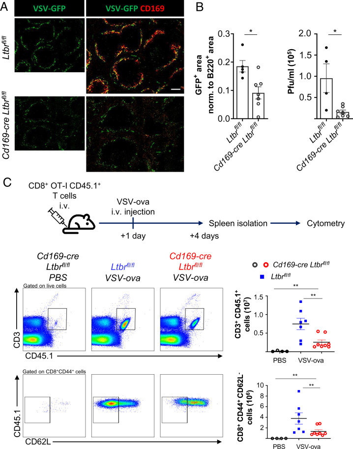
Impaired antiviral immunity in the absence of MMMs. (A) Wide-field immunofluorescence images for fluorescent VSV (VSV-GFP) and CD169 of spleens from Cd169-cre Ltbrfl/fl and Ltbrfl/fl mice. The graph depicts the mean area of GFP with individual data points normalized to the B220+ area. (B) The graph shows the quantification of the viral titer in the spleen of the indicated mice by measuring plaque-forming units. Shown are the mean with data points of individual mice. Statistical significance (Mann-Whitney test); *P < 0.05; error bar, SEM. (C) Experimental design for infecting mice with VSV-ova 24 h after bone marrow transfer of 5 × 10⁶ CD45.1 × OT-I mice. Gating strategy of flow cytometry analysis of ova-specific donor (CD45.1+ OT-I) T cells and their differentiation into cytotoxic CD8+ CD44+ CD62L− T cells 3 d after intravenous infection of ova-expressing VSV. Graphs depict the numbers of CD3+ CD45.1+ T cells (Top) and of CD8+ CD44+ CD62L− CD45.1+ T cells (Bottom). Shown are the mean values with data points of individual mice. Statistical significance (Mann-Whitney test); **P < 0.005; error bar, SEM (scale bar, 100 μm).

