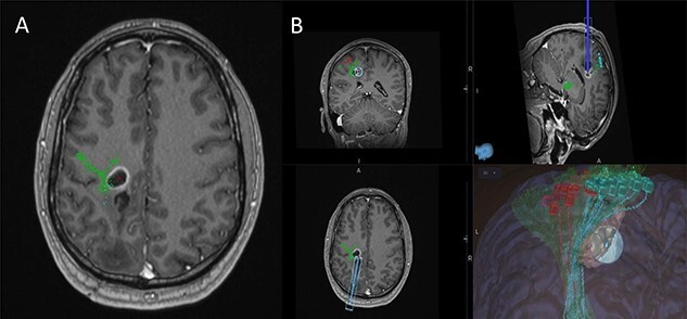
Figure 3 .

(A) Axial T1-weighted image with gadolinium showing the lesion with imposed DTI tractography of the CST; (B) planned trajectory for insertion of the tubular retractor guided by the preoperative integrated anatomical and functional motor mapping.
