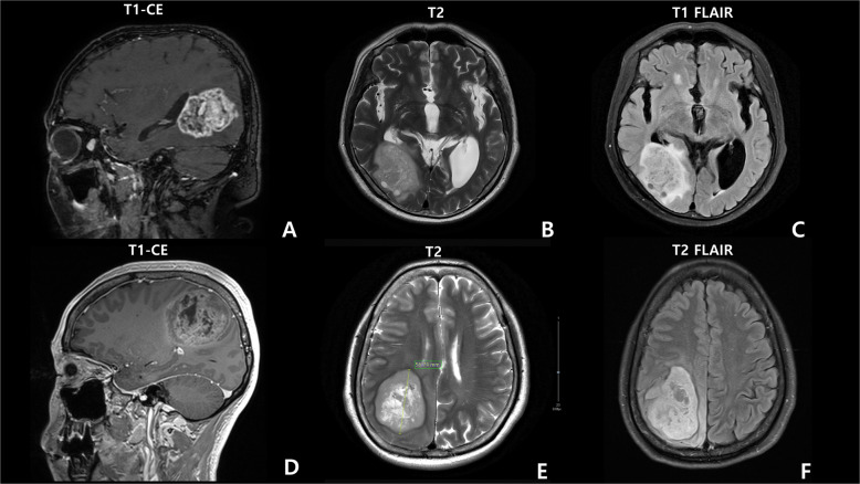
Fig. 2. The brain MRI images of case 4 with Lynch syndrome.
A sagittal T1-weighted (postcontrast), B axial T2-weighted, and C T2 FLAIR MRI results, showing an ~6 cm-long diameter enhancing mass with perilesional edema in the right occipital lobe. Case 2 (glioblastoma IDH-wildtype) D sagittal T1-weighted (postcontrast), E axial T2-weighted, and F T2 FLAIR MRI results, revealing an ~5.7 cm heterogeneous mass in the right parietal lobe and midline shift.

