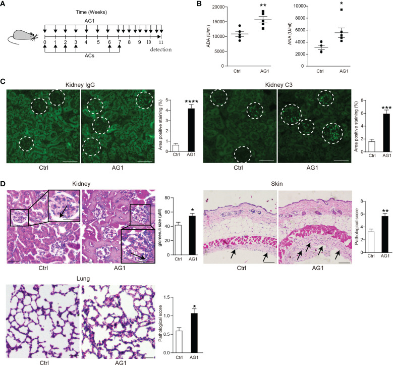
Figure 4.
Interfering the PPP activity regulates the process of SLE. (A) Eight-week-old female WT mice were intravenously injected with 1.5 × 10⁷ apoptotic thymocytes by four consecutive injections once a week; after 15 days, the injections were repeated twice. Meanwhile, 24 h before apoptotic cell injection, AG1(10 mg/kg, i.p.) was administered weekly (AG1 is still injected at a fixed time during the 15-day break). After the last apoptotic cells injection, AG1 was injected twice per week. The same volume of PBS was injected into the control group. (B) Serum concentrations of anti-dsDNA antibodies (ADA) (n = 5) and anti-nuclear antibodies (ANA) (n = 6) were detected by ELISA. (C) Representative images and percentages of positive staining areas of IgG and C3 in kidneys of AG1-treated SLE mice and untreated SLE mice (n = 5, bar = 50 μm). (D) Representative images and quantitative assessment of hematoxylin-and-eosin (HE) staining of the kidney (bar = 50 μm), skin (bar = 100 μm), and lungs (bar = 100 μm) from AG1-treated SLE mice and untreated SLE mice (n = 5 per group). *p < 0.05, **p < 0.01, ***p < 0.001, and ****p < 0.0001 (two-tailed Student’s t-test).

