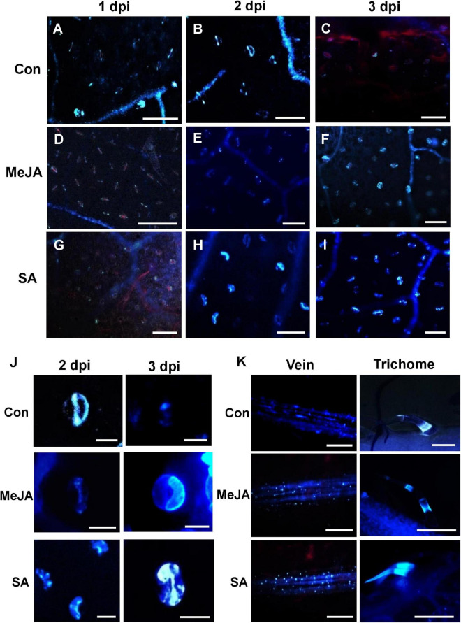
FIGURE 10.
Comparison of callose deposition on the leaf surface, veins, stomata, and trichomes show that SA priming enhanced callose deposition in response to infection in the early phases while MeJA enhanced in the later phases. (A–C) The control plants show early callose deposition on leaf surface, veins, and stomata over the first two days of infection, while on the third day callose is degraded due to necrosis. (D–F) Deposition of callose in the veins, stomatal guard cells in MeJA-primed leaves with more prominent deposition starting on the 3 dpi (G–I) Callose deposition in the SA-primed leaves started on the second day and was significantly more than that of MeJA-primed plants. Three images were analyzed from four individual replicates. Bar = 50 μm. (J) Enlarged view of the stomata at 2 and 3 dpi. In the control leaves, only one of the guard cells of each stoma fluoresced on 2 dpi and then by third day it diminishes. In MeJA, no fluorescence on 1–2 dpi but both guard cells fluorescence on 3 dpi. In SA-primed leaves, at 1 dpi there is no fluorescence, at 2 dpi only one guard cell fluorescence and in the following day both of the guard cells fluoresce. (K) Anniline blue fluorescence of the veins and trichomes at 2 dpi shows less callose deposition in the control compared to the phytohormone primes leaves. Bar = 300 μm.

