FIGURE 3.
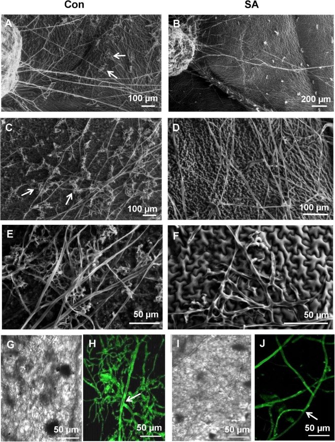
Scanning Electron Microscopy and Confocal Laser Scanning Microscopy (CLSM) to observe differential behavior of sclerotia and the newly emerging infection hyphae of R. solani transformed with gfp gene on SA-primed and control leaves up to 48 hpi. (A) SEM of germinating sclerotia showing profuse growth on the control leaf, presence of hyphal aggregation, and initiation of infection cushions (arrows) at 24 hpi. (B) Delayed germination of sclerotia with significantly less emerging hyphae and no infection cushion on the surface of SA-primed leaf at the same point (C) Numerous infection cushions on control leaf surface at 48 hpi (D) Hyphae growing in straight lines with scant hyphal branching and light hyphal aggregation at 48 hpi on SA pre-treated leaves. (E) SEM of close-up view of advanced infection cushion on the control leaf at 48 hpi (F) A close-up view of loose hyphal aggregation on SA-primed leaf at 48 hpi. (G,H) Confocal microscopy of gfp-transformed R. solani hyphae on control leaf surface at 24 hpi showing profuse branching, arrow showing right angled branching with septa typical for R. solani. Representative fluorescence fields were chosen from at least three independent plants. (I,J) Fluorescence microscopy of inoculated SA-primed leaves showing scant hyphal growth at the same time point (arrow shows right angle branching with septa typical for R. solani).

