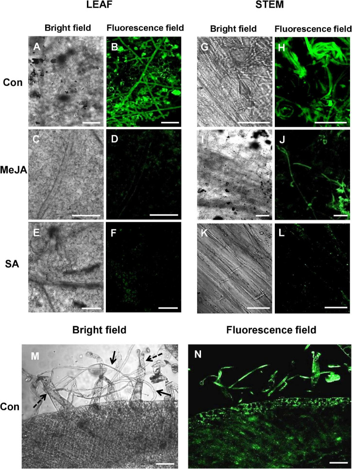
FIGURE 6.
Confocal Laser Scanning Microscopy (CLSM) of R. solani transformed with green fluorescence protein gene (gfp) on infected tomato leaves and stems showing avoidance of primed plants, especially SA-primed plants at 24 hpi. (A,B) CLSM of control tomato leaves showing profuse hyphal growth with infection cushions in close contact with the leaf surface. (C–F) JA and SA-primed leaves show significantly less growth of hyphae at 24 hpi. (G–L) Confocal microscopy of stems of primed and control plants infected with transformed R. solani showing least growth on SA-primed stem. (M,N) Transverse section of infected control stem showing significant interaction of hyphae (solid arrows) with stem-surface trichomes (dashed arrows) and infection within tissue at 24 hpi. Representative fluorescence fields were chosen from at least three independent plants. Bar = 50 μm.

