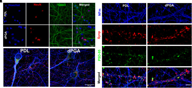
Figure 2.
Differentiation of primary neocortical neurons cultured on dPGA. (A) E18 rat neocortical neurons at 12 DIV on coverslips coated with 100 µg/ml of PDL or dPGA and immunolabled for neuronal markers NeuN and β3-Tubulin (tubb3, scale bar 20um). (B) E18 rat neocortical cultures at 12 DIV on coverslips coated with 100 µg/ml of either PDL or dPGA and stained with neuronal specific NFM, and synaptic markers Synaptophysin-1 (Synp) and PSD-95 (scale bar 20um). (C) E18 rat neocortical neurons at 12 DIV on coverslips coated with 100 µg/ml of PDL or dPGA and immunolabeled for NFM, presynaptic Synaptophysin-1 and postsynaptic PSD-95 (scale bar 2 µm).

