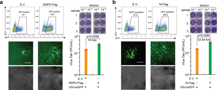
Fig. 2.
SARS-CoV-2 NSP5 and N facilitate viral replication. The HEK293T cells were subjected to transfection with empty vector (E.V.), NSP5 (a)-, or N (b)-expressing plasmids as indicated for 24 h. The cells were subsequently infected with VSV-eGFP (MOI = 0.001) for 12 h before imaging and flow cytometry analysis. The culture supernatant collected at 20 h postinfection was used to determine viral titers (PFU per mL) via plaque assays. The fluorescent imaging and flow cytometry data are representative of two independently performed experiments with similar results. Scale bar, 50 μm. In plaque assays, data are presented as mean values ± SEM from triplicate infections from one representative experiment of two. Statistical significance is shown as indicated. EV empty vector, h hours, PFU plaque-forming units.

