Figure 1.
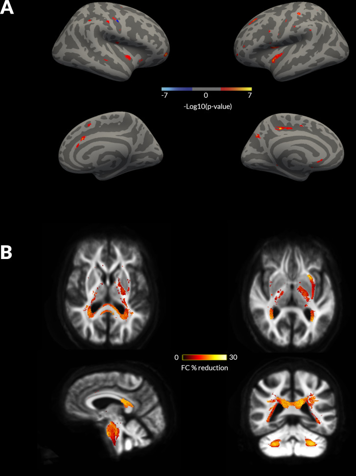
Whole brain, grey matter and white matter changes in patients with Parkinson’s disease (PD) and visual hallucinations (VH) at 18 months follow-up. (A) Changes in cortical thickness seen in patients with compared with those without hallucinations (PD non-VH) at 18 months follow-up, on a surface rendered right hemisphere (left: lateral view top panel, medial view bottom panel) and left hemisphere. No statistically significant changes were seen in cortical thickness at baseline imaging. Colour coding indicates cluster significance for group-by-time interactions. Significance levels are on a logarithmic scale of p values (−log10). Positive values indicate PD-VH cortical thickness <PD non-VH; negative values indicate PD-VH >PD non VH. Results are corrected for false discovery rate across both hemispheres. (B) Changes in white matter macrostructure (fibre cross-section, FC) seen in PD-VH compared with PD non-VH at longitudinal follow-up. Baseline changes are presented in online supplemental figure 1. Results are displayed as streamlines; these correspond to fixels that significantly differed between PD low and high visual performers (family-wise error correction (FWE)-corrected p<0.05). Streamlines are coloured by percentage reduction (colourbars) in PD-VH compared with PD non-VH.

