Figure 2.
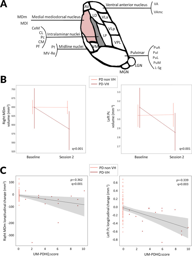
Thalamic subnucleus specific changes in PD with visual hallucinations compared with PD without hallucinations at 18 months follow-up. (A) Diagrammatic representation of the human thalamic subnuclei. Highlighted in pink are the nuclei showing significant longitudinal changes in volume in PD with visual hallucinations (PD-VH) compared with PD without hallucinations (PD non-VH). The medial mediodorsal nucleus is further subdivided into mediodorsal medial parvocellular and mediodorsal medial magnocellular nuclei. Intralaminar nuclei include the Pc, Pf, CeM, CL and CM. (B) Longitudinal change in thalamic nuclei volumes for the right MDm and the left Pc in PD-VH, PD nonVH in mm3. Corrected for age, gender, total intracranial volume and time between scan. Family wise error (FDR) corrected p-value presented for the group-by-time interaction comparison between PD-VH and PD non-VH participants. Error bars represent SD. (C) Change in thalamic nuclei volumes for the right MDm and the left Pc in PD participants was correlated with severity of visual hallucinations, assessed using the University of Miami Parkinson’s disease Hallucinations Questionnaire. Higher scores indicating more severe hallucinations. AV, anteroventral; CeM, central medial; Cl, central lateral; CM, centromedian; L_Sg, limitans; LD, laterodorsal; LGN, lateral geniculate; LP, lateral posterior; MDl, mediodorsal medial parvocellular; MDm, mediodorsal medial magnocellular; MGN, medial geniculate; MVRe, reuniens medial ventral; PD, Parkinson’s disease; Pf, parafascicular; PuA, pulvinar anterior; PuI, pulvinar inferior; PuL, pulvinar lateral; PuM, pulvinar medial; VA, ventral anterior; VAmc, ventral anterior magnocellular; VLa, ventral lateral anterior; VLp, ventral lateral posterior; VPL, ventral posterolateral; VM, ventromedial; ρ, Spearman correlation coefficient.

