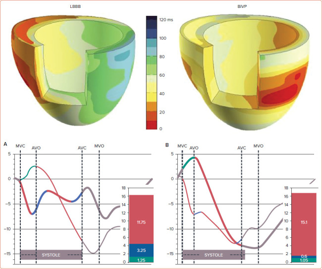
Figure 1: Electrical Activation and Contraction in Left Bundle Branch Block and Biventricular Pacing.

Upper panels: 3D representation of ventricular electrical activation in a canine heart with LBBB before (left) and during BiVP (right). Lower panels: strain patterns from a patient with LBBB before (left) and during BiVP (right). Thick tracings are septal strain; thin tracings are left ventricular free wall strain. Contribution of each of the following strains to total shortening is depicted in the coloured bars. On the basis of the slope of the deformation curve systolic deformation at each wall is divided into shortening (systolic total shortening; red), stretching preceded by shortening (SRS; blue), or early stretching not preceded by shortening (SPS; green). Systolic stretch index is calculated as SRS + SPS. AVC = aortic valve closure; AVO = aortic valve opening; BiVP = biventricular pacing; LBBB = left bundle branch block; MVC = mitral valve closure; MVO = mitral valve opening; SPS = systolic pre-stretch; SRS = systolic rebound stretch. Source: Upper panel: Strik et al. 2013.[55] Adapted with permission from Wolters Kluwer. Lower panel: De Boeck et al. 2009.[33] Adapted with permission from John Wiley & Sons.
