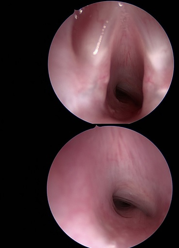
Figure 2.

Two intraoperative endoscopic images taken at MLB prior to excision of the cervical mass, at glottic level and in subglottis, showing partial airway obstruction secondary to extrinsic tracheal compression extending from subglottis to mid tracheal level. MLB, microlaryngoscopy and bronchoscopy.
