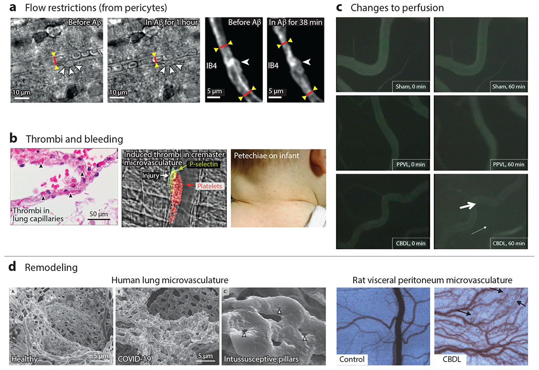
Figure 4.

Microvascular dysfunction in vivo occurs from flow restrictions, thrombi, bleeding, microvascular remodeling, and changes to perfusion. (a) Bright-field and two-photon fluorescence of rat cortical slices show constriction near pericytes. Less conspicuous, albeit significant, systemic 25% changes in diameter can cause reductions in blood flow on the order of 50% in the brain (39). (b) Microthrombi, especially when systemically distributed, can cause organ damage and death. Here, microthrombi are shown from the alveolar capillaries of a patient who died from COVID-19 (left) or induced by laser injury in murine cremaster microvasculature (middle). Petechiae are from capillary bleeding, seen here on an infant with viral illness (right). Panel b adapted from References 38 (left subpanel), 46 (middle subpanel), and 120 (right subpanel). (c) Common bile duct ligation causes changes to the vascular permeability as seen by the increased fluorescent signal in the interstitium. Panel c adapted from Reference 121. (d) Microvasculature can also remodel in response to pathological conditions, shown here for a COVID-19 patient. In particular, intussusceptive angiogenesis appears to be occurring as evidenced by the intussusceptive pillars. Changes to the microvascular density in the visceral peritoneum of rats can be seen with common bile duct ligation. Panel d adapted from Reference 121. Abbreviations: Aβ, amyloid beta; CBDL, common bile duct ligation; IB4, isolectin B4; PPVL, partial portal vein ligation.
