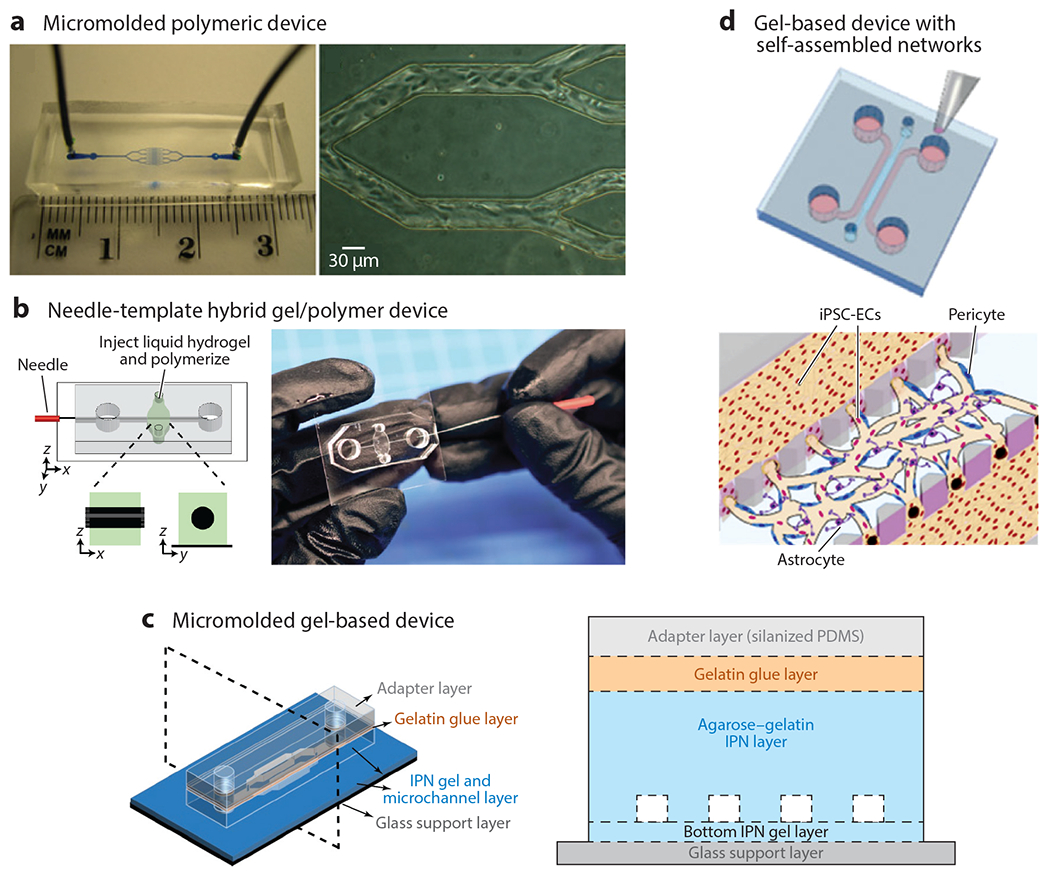
Figure 5.

Approaches to creating in vitro microvasculature can be categorized as using polymeric or very soft gel materials with template-based, self-assembled, or directed networks of endothelial cells. (a) Polymeric-based micromolded structures offer high control over spatial dimensions and typically have a fast time to confluency. Panel a adapted from Reference 76. (b) Needle-templated hybrid structures offer key advantages of convenience and ease of fabrication, while using soft gels for perfusion studies, but typically are larger than most microvasculature (>100 μm). Panel b adapted from Reference 59. (c) Stiffer gels cleverly bonded with adapter layers led to the creation of the smallest in vitro vessels (~20 μm) that were still in a soft gel material for perfusion studies (35). (d) Pillars separating three fluidic channels enable gels laden with endothelial cells, pericytes, and astrocytes to be cast in the center gel and have perfusion in the adjacent channels to support culture and subsequent seeding after vascularization. Panel d adapted from Reference 75. Abbreviations: IPN, interpenetrating polymer network; iPSC-EC, induced pluripotent stem cell–derived endothelial cell; PDMS, polydimethylsiloxane.
