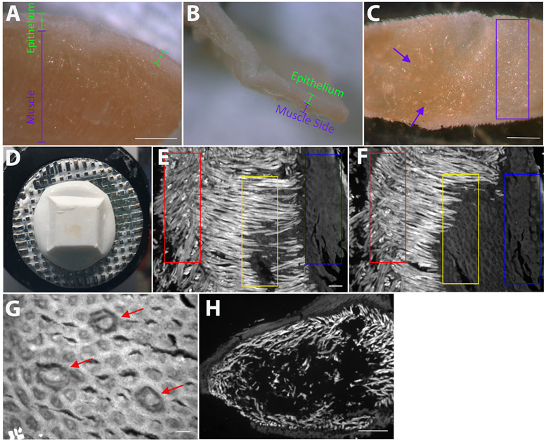
Figure 1: Preparation of lingual epithelium for fungiform taste-bud staining.
(A) View of the cut tongue with epithelium and muscle labeled prior to any dissection. (B) Once enough muscle has been removed, there is only a small amount of remaining muscle on the underside of the epithelium. In addition to evaluating the progress of the dissection by viewing the cut side of the epithelium, (C) laying the epithelium flat on a glass slide under the dissecting scope reveals that some portions of the tissue are evenly translucent (purple rectangle); enough muscle has been removed from this area. In contrast, the purple arrows indicate regions on the left where there is more muscle that needs to be removed. Once the entire underside of the epithelium is similar to the area in the purple rectangle, proceed to the next step. (D) After portions of the epithelium have been frozen with the muscle side down, additional muscle and lamina propria are removed as thin sections using the cryostat. When sectioning is complete, the remaining epithelium is thin and translucent. (E-F) Serial sections (20 μm) were collected on a glass slide, and each section was viewed under a fluorescent microscope before cutting the next section. Well below the epithelium, muscle fibers are oriented in multiple directions so that muscle fibers are present both in cross section and along the muscle fiber (E, red rectangle). The serial sections in E-F demonstrate the transition from muscle fibers oriented in multiple directions (E, red rectangle) to muscle fibers being oriented mostly in one direction (F, red rectangle), which is indicative of the muscle-lamina propria border. Another region of the same piece of tissue (yellow rectangles) demonstrates that when the muscle fibers are oriented in one direction, the next section will likely yield connective tissue because all muscle has been removed from that region. The blue rectangles both represent the underside of the epithelium. If taste buds are present on the section (G, red arrows), too much tissue has been removed. Ideally, sectioning is complete when the underside of the epithelium (but no taste buds) is visible in the removed sections (F, yellow rectangle). Although areas with muscle fibers oriented in the same direction (E, yellow rectangle and F, red rectangle) are also suitable for sectioning, areas where the muscle fibers are oriented in multiple directions (E, red rectangle) should be avoided. (G) Once sections include the underside of the epithelium/lamina propria, it is only possible to cut a few additional sections before too much of the epithelium has been removed and sections include taste buds. (H) The most common mistake is revealed by cryostat sections where epithelium is seen at the edge of the tissue, muscle is seen inside of the epithelium, and OTC/sparse muscle is present in the middle. This is most often due to not laying the tissue flat on the bottom of the tissue mold before freezing or insufficient flattening with blunt-ended forceps. Scale bars in A-C = 1 mm; scale bars in E, F, H = 100 μm; scale bar in G = 50 μm.

