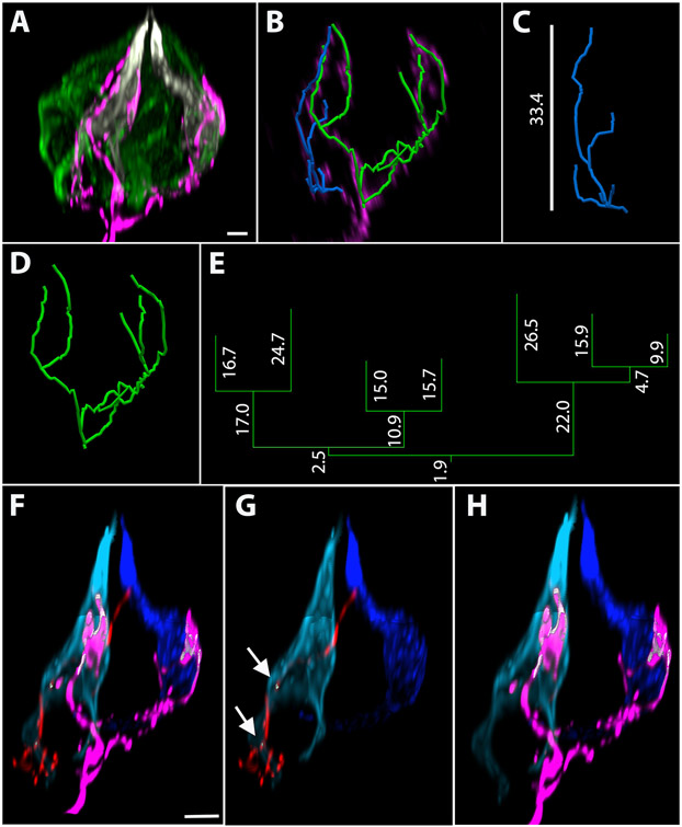
Figure 4: Representative terminal arbors in fungiform taste buds using sparse cell genetic labeling.
(A) Whole-mount taste bud stained with taste-transducing-cell markers Car4 (white) and PLCβ2 (green). (B) This taste bud has two labeled terminal arbors, which are shown with the taste bud removed after reconstructing the fibers. (C) The blue arbor has 6 branch ends and an orthogonal height in the taste bud of 33.4 μm and (D) the green arbor has 7 branch ends. (E) The dendrogram corresponding to the green arbor is provided with each segment length in micrometers. (F-H) The distance between structures was measured. (F-G) The blue tracing in C was segmented and is shown in red. (G) The areas where this terminal arbor is within 200 nm of the light blue Car4+ cell are indicated by white arrows. (F, H) The terminal arbor represented by the green reconstruction is shown in magenta. (H) The magenta arbor (associated with the green tracing in 4B, D) is within 200 nm of both the dark and light blue Car4+ cells. Scale bar in A, B = 4 μm; scale bars in F-H = 5 μm.

