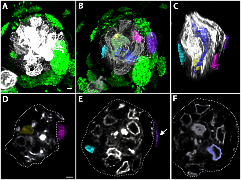
Figure 5: Whole-mounts can be used to track incorporation of new taste bud cells.
Mice were injected with EdU to label dividing progenitors on Days 0, 1, and 3 and sacrificed on Day 4. (A, B) Cells labeled with EdU (green) can be identified both around and within the taste bud, which is labeled with keratin-8 (A, white, B, gray). (B, C) Individual EdU-labeled, keratin-8+ cells inside the taste bud and keratin-8−, EdU-labeled nuclei are segmented outside the taste bud. (D-F) The fluorescence within each structure segmented in A-B was masked and can be seen in cross-section. The perimeter of the taste bud is outlined with a white dotted line (D-F). (D) The yellow cell is within the taste bud and is both EdU-labeled and keratin-8+. The magenta nucleus is outside the taste bud and is keratin-8−. (E) The teal cell is inside the taste bud and both EdU-labeled and keratin-8+. The purple EdU-labeled nucleus is keratin-8− and outside of the taste bud (white arrow). (F) The blue cell is keratin-8+ and elongated, consistent with mature taste-transducing cells. Scale bars in A-C =3 μm; scale bars in D = 2 μm; scale bars in E, F = 4 μm. Abbreviation: EdU = 5-ethynyl-2′-deoxyuridine.

