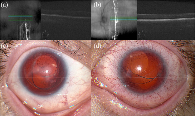
Figure 3.
Spectral-domain optical coherence tomography (SD-OCT) and anterior segment photography of a 29-year-old male with aniridia, nystagmus, bilateral limbal stem cell deficiency, and pseudophakia, and a best corrected visual acuity of 20/400 in the right eye and 20/200 in the left eye. SD-OCT of (a) right and (b) left eyes is technically suboptimal due to nystagmus and inability to fixate, but shows blunted foveal contours. Anterior segment photography of the (c) right and (d) left eyes shows aniridia, corneal neovascularization, and decentered in-the-bag posterior chamber intraocular lenses.

