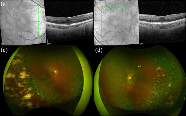
Figure 7.
Spectral-domain optical coherence tomography (SD-OCT) and fundus photography of a 14-year-old female with history of bilateral aggressive posterior retinopathy of prematurity treated with laser, cerebral infarction, strabismus, and a best corrected visual acuity of 20/30 in the right eye and 20/50 in the left eye. SD-OCT of (a) right and (b) left eyes shows blunted foveal contours. Fundus photography of (c) right and (d) left eyes shows blunted foveal reflex, temporal vessel straightening, and retinal laser ablation.

