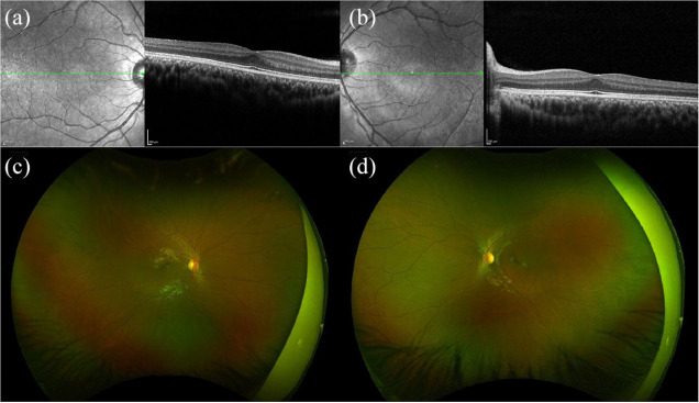
Figure 8.
Spectral-domain optical coherence tomography (SD-OCT) and fundus photography of a 16-year-old male with autism and a best corrected visual acuity of 20/20 bilaterally. SD-OCT of (a) right and (b) left eyes shows blunted foveal countours. Fundus photography of (c) right and (d) left eyes shows blunted foveal reflexes.

