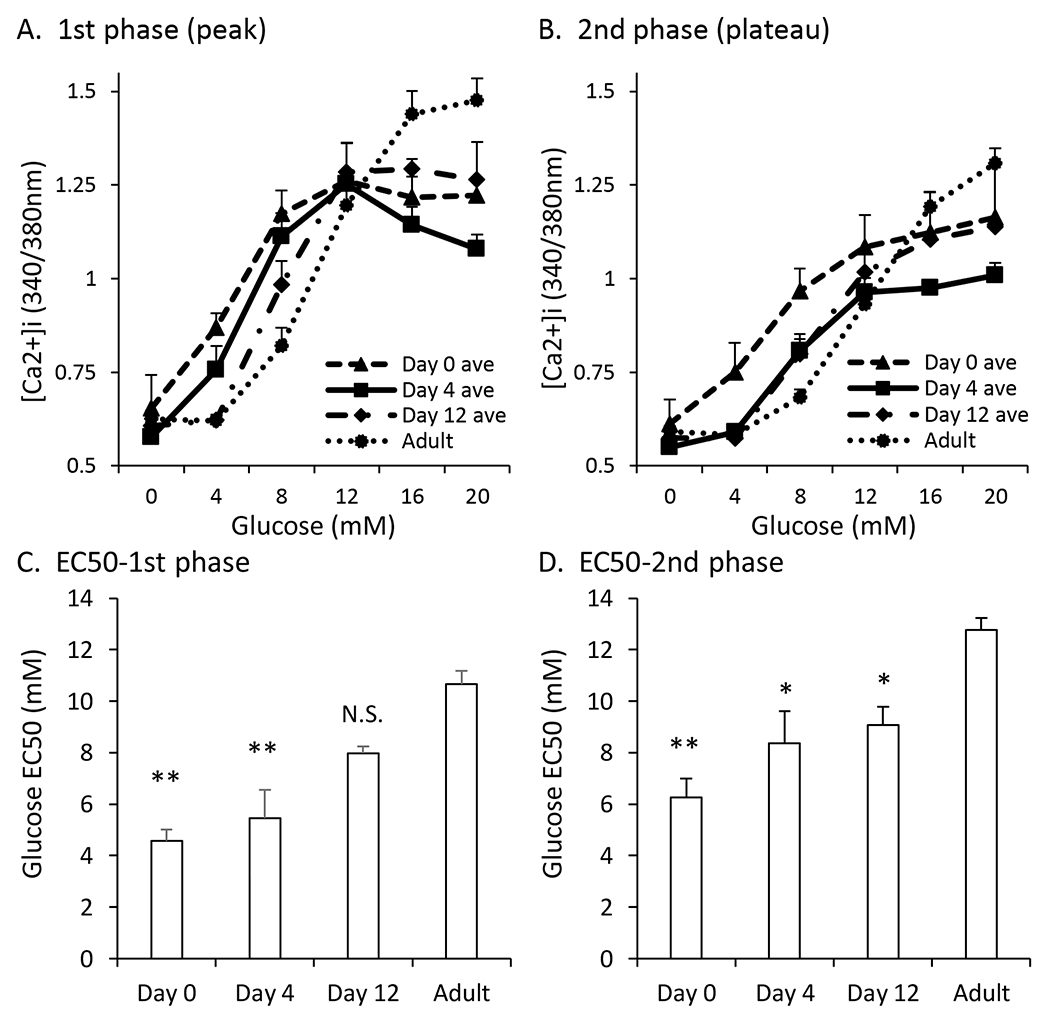
Figure 2. Combined glucose curves show left-shifted pattern.

A-B. Traces showing 1st phase peak (A) and 2nd phase plateau (B) [Ca2+]i responses to glucose stimulation from 0 to 4 to 8 to 12 to 16 to 20mM for islets aged postnatal day 0 , day 4, day 12, and adult (dotted black). C-D. EC50 values of the maximum peak (C) and plateau (D) of islets at postnatal day 0, day 4, day 12, and adult. Asterisks (*) indicate differences between adult and neonatal [Ca2+]i traces based on N=3 sets of islets for each neonatal day and N=9 for control. *P<0.05, **P<0.01, N.S. Not significant.
