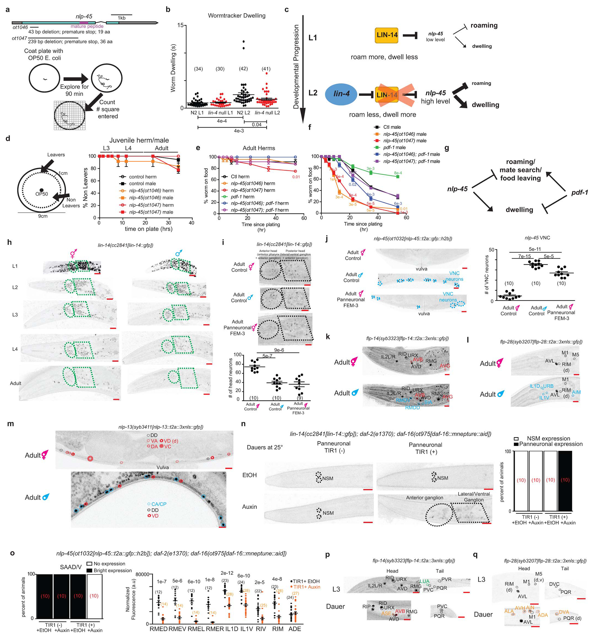
Extended Data Fig 10. Regulation and function of nlp-45 across temporal, sexual, and environmental dimensions of post-embryonic development.

a, Schematic of nlp-45 deletion mutants and exploratory assay. b, Increased dwelling during L1->L2 transition is partially juvenized in lin-4(e912) animals. Mean +/− SEM and n (in bracket) are shown for each condition, and each point of the scatter dot plot represents a single animal. Wilcoxon rank-sum tests and false-discovery rate q values for each comparison shown below. c, Schematic showing lin-4/lin-14 regulation of nlp-45 to alter exploratory behavior during the L1>L2 transition. d, nlp-45 deletion mutants do not significantly affect the food leaving behaviors of juvenile males/hermaphrodites. Values were plotted as mean +/− SEM of three independent experiments (n=6 animals per independent experiments). e, Leaving assay for adult hermaphrodite in nlp-45 and pdf-1 mutant animals. Values were plotted as mean +/− SEM of three independent experiments (n= 8 animals per independent experiments). Statistical analysis (post-hoc two-sided t-test) is only shown for the comparison to respective controls (color coded respectively). f, Leaving assay for adult male in nlp-45 and pdf-1 mutant animals. Values plotted as mean +/− SEM of three independent experiments (n= 8 animals per independent experiments). Statistical analysis (post-hoc two-sided t-test) is only shown for the comparison to respective controls (color coded respectively). g, Schematic of the opposing role of nlp-45 and pdf-1 on male food leaving behavior. h, Developmental expression of lin-14 in hermaphrodites and males. Representative images for the lin-14(cc2841[lin-14::gfp]) reporter are shown across all developmental stages for both sexes. LIN-14 expression was similarly downregulated in both sexes at early larval stages, its expression in the late larval and particularly in the adult stage was significantly more reduced in the male nervous system compared to that of the hermaphrodite. Ellipse and polygon outline anterior and lateral/ventral neuronal ganglia. Representative images for hermaphrodite are re-used here from Extended Data Fig.5b for direct side by side comparison with male animals across development. i, Panneuronal depletion of sex determination master regulator TRA-1, through overexpression of FEM-3, decreases nervous system LIN-14 expression in adult hermaphrodites to mimic that of adult males. Representative microscope images, shown above, are overexposed in comparison to previous lin-14 reporter images to better show the dim expression in adult males and FEM-3 overexpressed hermaphrodites. The quantifications of head neuron numbers across the three conditions are shown below. The mean +/− SEM and n (in bracket) shown for each condition, and each point of the scatter dot plot represents a single animal. Post-hoc two-sided t-test p values are shown for each comparison. j, Panneuronal depletion of TRA-1, through overexpression of FEM-3, masculinizes nlp-45 expression in adult hermaphrodite VNC. Representative images are shown on the left. Quantifications of VNC neuron numbers are shown on the right. The mean +/− SEM and n (in bracket) are shown for each condition, and each point of the scatter dot plot represents a single animal. Post-hoc two-sided t-test p values are shown for each comparison. k, Sexually dimorphic expression of flp-14 in adult hermaphrodites and males. In addition to stronger flp-14 expression in the AVB neuron (red) as compared to adult hermaphrodites, adult males gain flp-14 expression in the RMDD/V and SIA neurons (blue). Adult hermaphrodite images are re-used from Extended Data Fig.3i. l, Sexually dimorphic expression of flp-28 in adult hermaphrodites and males. As compared to adult hermaphrodites, adult males gain flp-28 expression in the IL1D/V, URB, and AIM neurons (blue). Adult hermaphrodite images are re-used from Extended Data Fig.3f. m, Sexually dimorphic expression of nlp-13 in adult hermaphrodites and males. In addition to stronger nlp-13 expression in the VD neurons (red) as compared to adult hermaphrodites, adult males gain nlp-13 expression in the male specific CA/CP motor neurons (blue) and lose nlp-13 expression in the DA, VA and hermaphrodite specific VC motor neurons. Adult hermaphrodite images are re-used from Extended Data Fig.3j. n, Panneuronal degradation of DAF-16 in auxin-treated dauers leads to panneuronal de-repression of lin-14 in dauer animals. Representative images are on the left while binary quantifications of panneuronal expression are shown on the right (number of animals for each condition is shown in red brackets). Animals were grown (from embryo onward) on NGM plates supplemented with OP50 and 4mM auxin in EtOH (indole-3 acetic acid, IAA, Alfa Aesar) at 25°C to degrade DAF-16 panneuronally and to induce dauer formation. As controls, plates were supplemented with the solvent EtOH instead of auxin. Additional control animals without panneuronal TIR-1 expression grown on EtOH and auxin were also included for comparison. o, Panneuronal degradation of DAF-16 in auxin-treated dauers leads to a loss or reduced nlp-45 expression in several neuronal classes. Representative images are shown in Fig. 4f. Binary quantifications are shown for the SAAD/V neurons (number of animals for each condition is shown in red brackets) while fluorescence quantifications are shown for the RMED/V, RMEL/R, IL1D/V, RIV, RIM, and ADE neurons. The mean +/− SEM and n (in bracket) are shown for each condition, and each point of the scatter dot plot represents a single animal. Post hoc two-sided t-test p values are shown for each comparison. p, Expression of flp-14 in dauer animals as compared to L3 animals. Upon entry into dauer, similar to expression pattern in the adult hermaphrodite/male, animals gain flp-14 expression in the AVB neurons (red) and lose flp-14 expression in the LUA neurons (green). Additional dauer specific flp-14 expression is gained in the ASE neurons (orange). Expression in the PVR neurons is also lost in dauer animals. L3 hermaphrodite images are re-used from Extended Data Fig.3i. q, Expression of flp-28 in dauer animals as compared to L3 animals. Upon entry into dauer, animals gain flp-28 expression in the ALA, AVH, AIN, ADA, and DVA neurons (labelled in orange) and lose flp-28 expression in the DVC neurons. L3 hermaphrodite images are re-used from Extended Data Fig.3f. d and v in brackets denote dim and variable expression, respectively. Scale bars = 10μm.
