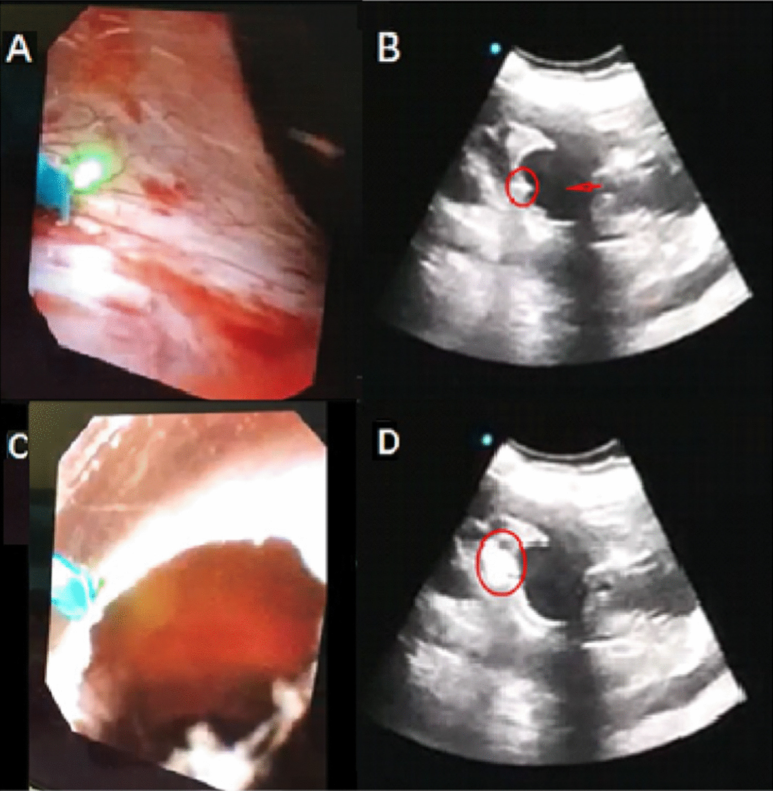
Fig. 1.

Surgical procedures of ultrasound-guided flexible ureteroscopy. A The operator approached the wall of the suspected cyst under a flexible ureteroscopic view and palpated the mucosa in this area. B Assistants applied ultrasound to monitor ureteral flexible scopes for contact and compression of the cyst (the arrow points to the cyst, and the rounded area is the end of the flexible ureteroscope). C The operator used a laser to cut in the area where the cyst was identified. D Assistants used ultrasound to monitor the incision of a parapelvic cyst (the circular area has a smoky appearance on the ultrasound image when the laser was working.)
