Fig. 3.
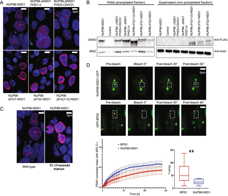
NUP98 FG repeat domains mediate the formation of NUP98-NSD1 liquid-like phase separated nuclear condensates. A Immunofluorescence staining of HEK293 cells transiently overexpressing full-length NUP98-NSD1 and its mutated forms. The cells were stained with Anti-FLAG antibody labeled with Alexa Fluor 568 secondary antibody (red). Nuclei were counterstained with DAPI (blue). B Precipitation of NUP98-NSD1 and its mutated forms in the presence of 30 μM b-isoxazole. Deletion of one or both FG repeat domains causes a decrease in the phase transition capacity of the fusion protein, represented by the signal in the supernatant. C Immunofluorescence analysis of HEK293 cells upon the treatment with 3% 1,6 hexanediol. The cells were stained with Anti-FLAG antibody labeled with Alexa Fluor 568 secondary antibody (red). Nuclei were counterstained with DAPI (blue). NUP98-NSD1 nuclear puncta were dispersed upon the treatment. D) Representative images of FRAP experiment of NUP98-NSD1-GFP and GFP-BP53 expressing HEK293 cells, with white boxes highlighting the punctum undergoing targeted bleaching. Fluorescence recovery over time was followed and quantified for NUP98-NSD1-GFP and GFP-BP53 (lower left panel). T-test comparison of the acquired fluorescence recovery half-times (T-half) was performed showing statistically significant faster recovery half-time for NUP98-NSD1-GFP. **P < 0.01, N = 54

