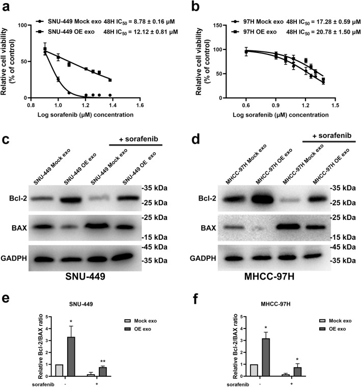
Fig. 3.
Exosomes from GOLPH3-overexpressed cells enhanced sorafenib resistance in HCC cells. a IC50 value of SNU-449 treated with indicated exosomes from 3 independent experiments. b IC50 value of MHCC-97H treated with indicated exosomes from 3 independent experiments. c For SNU-449 cells incubated with indicated exosomes and thereafter treated with sorafenib (10 μmol/L) for 48 h, Bcl-2 and Bax levels were determined by western blot, with GAPDH as the normalized control. d For MHCC-97H cells incubated with indicated exosomes and exposed to 20 μmol/L sorafenib for 48 h, levels of Bcl-2 and Bax were detected via western blotting. e, f Grayscale value analysis of Bcl-2/Bax ratio by Image J. Error bars represent mean ± SD from 3 independent experiments. Data are analyzed by t test (a, b) or two-way ANOVA with Tukey’s multiple comparisons test (e, f). *p < 0.05; **p < 0.01

