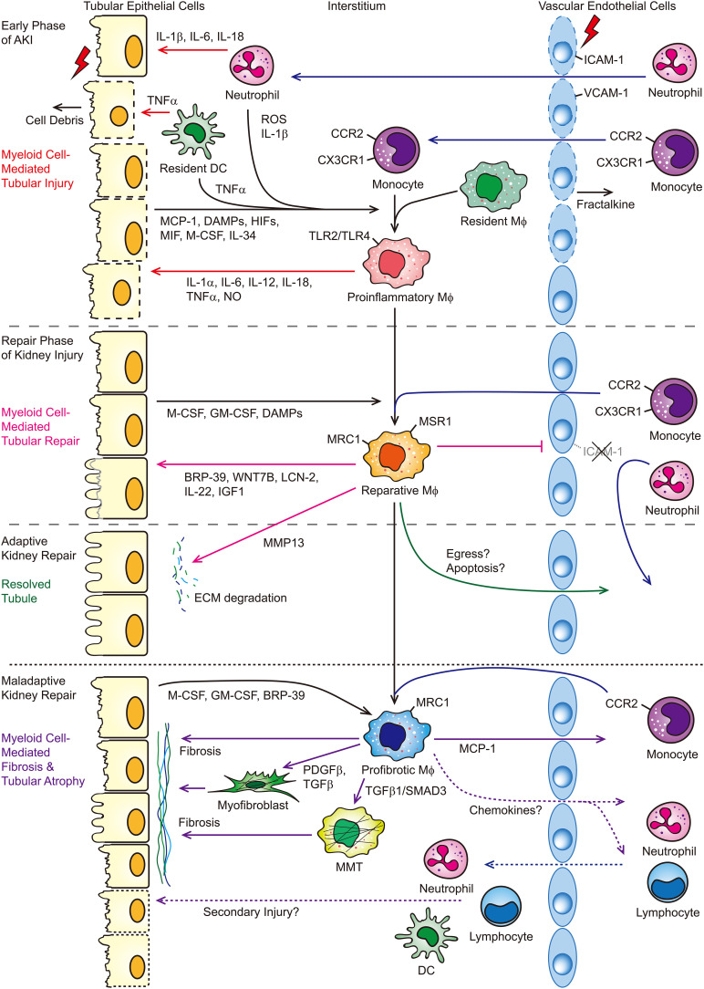
Figure 1.
Myeloid cells mediate tubular injury and repair following AKI. In response to AKI, endothelial cells express fractalkine (CX3CL1) and ICAM-1 and VCAM-1 to recruit neutrophils and monocytes into the injured kidney and promote leukocyte-endothelial cell adhesion, respectively. IL-1β, IL-6, IL-18, and TNF-α released from neutrophils/dendritic cells induce apoptosis and/or necrosis of tubular epithelial cells. Proinflammatory cytokines, ROS, HIFs, and DAMPs released by degranulating PMNs and injured or dying tubular cells lead to the proinflammatory activation of macrophages. These proinflammatory macrophages then subsequently produce proinflammatory cytokines and NO, which lead to further tubular injury. The inflammatory milieu in the early phase is later reversed during the repair phase. Downregulation of ICAM-1 and VCAM-1 limits inflammatory cell infiltration. Macrophage growth and differentiation factors M-CSF and GM-CSF and DAMPs released from the tubular epithelial cells lead to the reparative activation of macrophages. BRP-39, WNT7B, LCN2, IL-22, and IGF1 secreted from reparative macrophages promote tubular cell proliferation and/or repair. In the setting of adaptive kidney repair, macrophages release MMP13 to facilitate degradation of ECM, which is initially deposited after injury to aid repair, and macrophage egress and/or apoptosis after the injury is resolved. However, in the setting of maladaptive kidney repair, macrophages persist within the kidney. Macrophage growth factors, M-CSF, GM-CSF, and BRP-39 released from sustained injured tubular epithelial cells promote further recruitment and/or retention of macrophages and polarize macrophages into a profibrotic phenotype. Profibrotic macrophages or MMTs can promote kidney fibrosis directly and indirectly by activating interstitial myofibroblasts and contribute to the secondary tubular injury. BRP-39, breast regression protein 39; DAMP, damage-associated molecular pattern; DC, dendritic cell; ECM, extracellular matrix; HIFs, hypoxia-inducible factors; ICAM-1, intracellular adhesion molecule 1; LCN-2, lipocalin-2; Mϕ, macrophage; MCP-1, monocyte chemoattractant protein-1; M-CSF, macrophage colony-stimulating factor; MIF, macrophage migration inhibitory factor; MMP13, matrix metallopeptidase 13; MMT, macrophage-to-myofibroblast transition; MRC1, mannose receptor 1; MSR1, macrophage scavenger receptor 1; NO, nitric oxide; PDGFβ, platelet-derived growth factor subunit B; ROS, reactive oxygen species; SMAD3, mothers against decapentaplegic homolog 3; TLR, Toll-like receptor; VCAM-1, vascular cell adhesion molecule-1, WNT7B, Wnt family member 7B.

