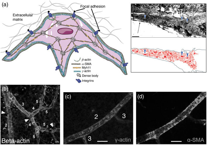
Fig. 3.
Putative organization of actin filaments in pericytes. The diagram is based on the available pericyte data and is inspired by extensive data from SMCs. - and -cytoplasmic actins are concentrated in cortical/submembranous regions, whereas -cytoplasmic actin is also located around dense bodies. -SMA, along with myosin filaments, stretches between focal adhesion points and dense bodies, forming the contractile stress fibers. This figure is produced using Servier medical art (http://www.servier.com). Inset shows a transmission electron micrograph of a cultured retinal pericyte process, harboring actin filaments and dense bodies in cytoplasm (arrowheads). Arrow indicates the extracellular matrix elements. Microfilaments and dense bodies are hand-outlined in red to depict their organization inside the pericyte process. Inset adapted with modifications from Schor et al.78 (b) Immunolabeling for cytoskeletal -actin, which runs along the cortex of the vessel in addition to diffuse labeling. Scale bar: . Adapted from Kureli et al.25 (c) and (d) - cytoplasmic actin and -SMA distribution in vessels after intravitreally injected phalloidin. Despite phalloidin stabilization of F-actin filaments, gamma-actin remained detectable only on ≤fourth-order branches unlike α-SMA. Scale bar: . (c) and (d) Adapted from Alarcon-Martinez et al.,23 with permission.

