Fig. 5.
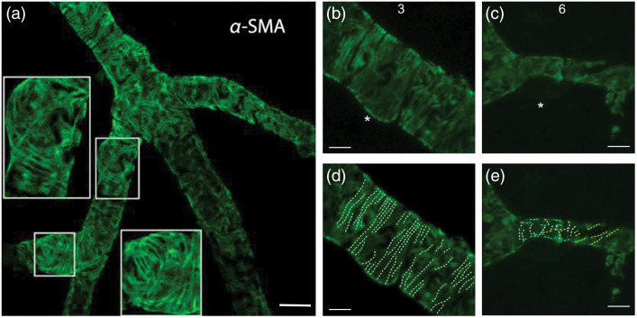
(a) -SMA fiber bundles form circular string-like structures reminiscent of contractile stress fiber organization. Insets: 2× magnified. (b)–(e) Bundles are differentially organized in pericytes from third and sixth retinal vascular orders. The organization of the -SMA labeling within the cytoplasm of pericyte processes suggest a vectoral structure, functioning in capillary diameter changes. It is noteworthy that in the (b) and (d) third order, bundles regularly run rather circumferential (white), while in the (c) and (e) sixth-order bundle orientation becomes irregular with some bundles running oblique (yellow) or parallel (cyan) to the longitudinal capillary axis. Scale bar: in (a), in (b-e).

