
FIGURE 3.
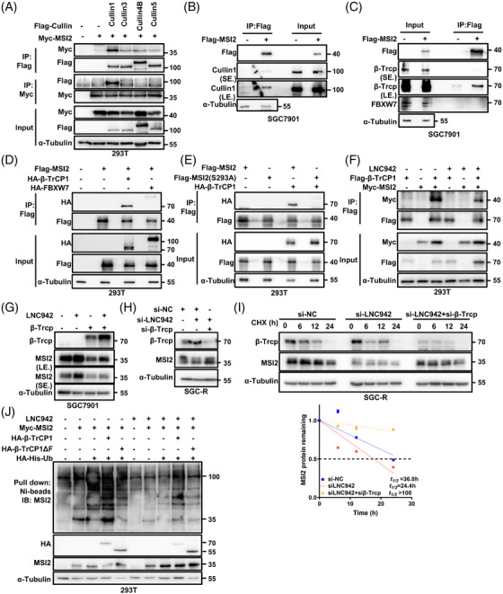
LNC942 disrupts the interactions between MSI2 and the SCFβ‐TRCP E3 ubiquitin ligase. (A) 293T cells were transfected with empty vector or Flag–Cullin family proteins and Myc‐MSI2 plasmids as indicated, followed by immunoprecipitation (IP) with anti‐Flag or anti‐Myc antibody and western blotting with the indicated antibodies. α‐Tubulin served as the loading control. After 48 h of transfection, the cells were treated with MG‐132 (20 μM) for 6 h before harvesting. (B) IP with anti‐Flag antibody was performed in SGC7901 cells transfected with empty vector or Flag–MSI2 plasmids. Western blotting was performed with the indicated antibodies. The cells were treated with MG‐132 (20 μM) for 6 h before harvesting. (C) Western blotting of input and anti‐Flag IP derived from SGC7901 cells transfected with Flag‐MSI2 plasmids. The cells were treated with MG‐132 (20 μM) for 6 h before harvesting. (D) Western blotting analysis of input and anti‐Flag IP derived from 293T cells transfected with Flag‐MSI2, HA‐β‐Trcp or HA‐FBXW7 plasmids. The cells were treated with MG‐132 (20 μM) for 6 h before harvesting. (E) Western blotting analysis of input and anti‐Flag IP derived from 293T cells transfected with HA‐β‐Trcp and the indicated Flag‐tagged MSI2 constructs. The cells were treated with MG‐132 (20 μM) for 6 h before harvesting. (F) IP was performed with anti‐Flag antibody in 293T cells transfected with Flag‐β‐Trcp and Myc‐MSI2 plasmids in the presence or absence of LNC942, followed by western blotting with the indicated antibodies. The cells were treated with MG‐132 (20 μM) for 6 h before harvesting. (G) The SGC7901 cells were co‐transfected with LNC942 and β‐Trcp plasmids, followed by western blotting of the MSI2 and β‐Trcp expression. (H) The SGC‐R cells were co‐transfected with LNC942 and β‐Trcp siRNAs, followed by western blotting of the MSI2 and β‐Trcp expression. (I) Western blotting detection of MSI2 protein half‐life in SGC‐R cells transfected with the indicated siRNAs and treated with cycloheximide (CHX) (100 μg/ml). The MSI2 protein abundance was quantified using ImageJ in the bottom panel. (J) 293T cells were co‐transfected with the indicated plasmids and treated with MG‐132 (20 μM) for 6 h before collection. MSI2 pull‐down experiments were conducted with nickel‐nitrilotriacetic acid (Ni‐NTA) beads, and the samples were analysed using western blotting with the indicated antibodies. Ub, ubiquitin. Data in (I) are shown as mean ± SD of three independent experiments