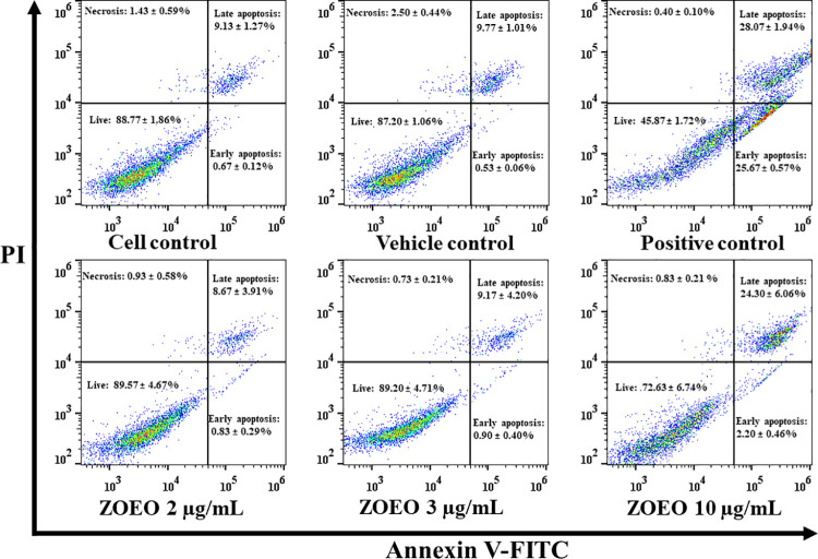
Fig 5. Apoptosis assay by flow cytometry after staining with double annexin V-FITC/propidium iodide (PI).
MCF-7 cells were treated with ZOEO at the concentrations of 2, 3, and 10 μg/mL. Representative flow cytometry dot plot indicating the cell population in apoptotic and necrotic quadrants after treatment with various concentrations. The dataset is available in S7 Table.

