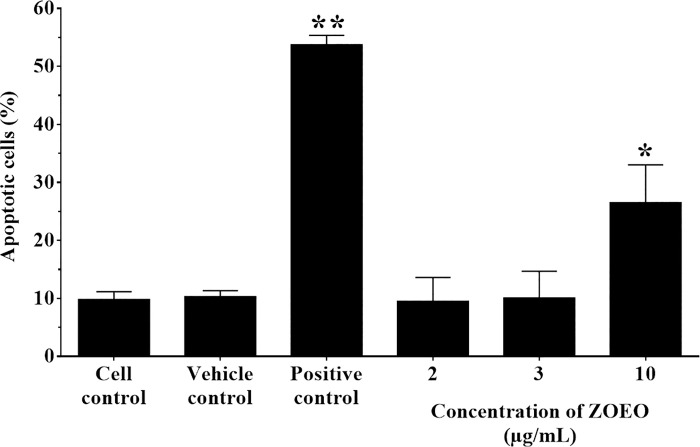
Fig 6. Representative bar graph of apoptotic cells (%) by flow cytometer after ZOEO treatments in various concentrations.
MCF-7 cells were treated with ZOEO at the concentrations of 2, 3, and 10 μg/mL and stained with double annexin V-FITC/propidium iodide (PI). The percentage of apoptotic cells was statistically compared. Each bar represents mean ± SD of three independent experiments performed in triplicate. Asterisk (*) denotes significant differences from vehicle control; * p < 0.05, ** p < 0.001. The dataset is available in S7 Table.

