
Figure 2.
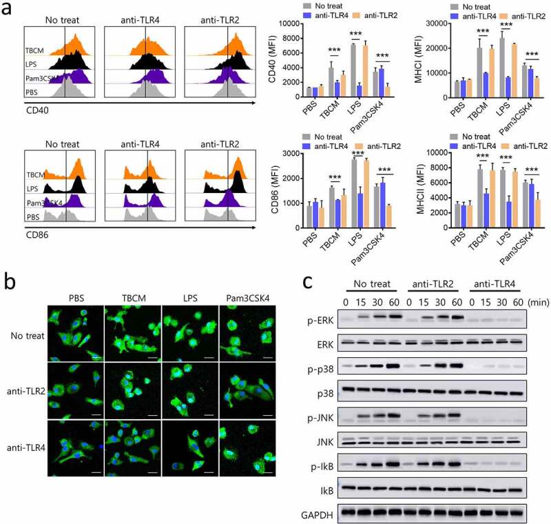
TBCM induces DC activation via activation of MAPKs and NF-kB in DCs in a TLR4–dependent manner. BMDCs were incubated with anti-TLR2 IgG or anti-TLR4 IgG prior to incubation with TBCM (1 μg/ml), LPS (0.1 μg/ml) or Pam3CSK4 (0.1 μg/ml). (a) After 24 h incubation with TBCM, LPS or Pam3CSK4, the surface expression of CD40, CD86, MHC class I and MHC class II was assessed by flow cytometry. (b) The effects of TBCM on the cellular localization of the p65 subunit of NF-kB in DCs were assessed. After 12 h of stimulation with TBCM, LPS or Pam3CSK4, the intracellular localization of NF-kB p65 was determined by immunofluorescence. (c) DCs were harvested at the indicated time points, and then the DC lysates were subjected to SDS-polyacrylamide gel and an immunoblot analysis was conducted. Significant differences (*p 0.05, **p 0.01, and ***p 0.001) among the different groups are shown in the related figures, and the data are presented as the means s.e.m. of three independent experiments.