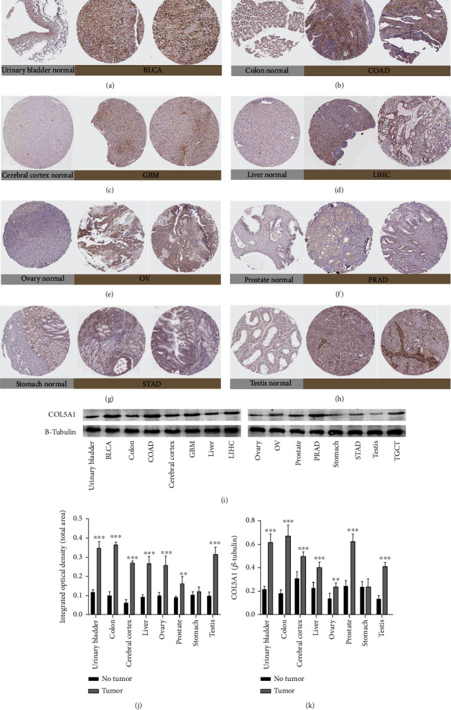
Figure 2.

Representative photographs of immunohistochemical staining and western blots of different normal tissues (left panels) and tumor tissues (right panels). The level of the COL5A1 protein was increased in bladder urothelial carcinoma (BLCA), colon adenocarcinoma (COAD), glioblastoma multiforme (GBM), liver hepatocellular carcinoma (LIHC), ovarian cancer (OV), prostate adenocarcinoma (PRAD), and testicular germ cell tumors (TGCTs). (a) Urinary bladder. (b) Colon. (c) Cerebral cortex. (d) Liver. (e) Ovary. (f) Prostate. (g) Stomach. (i) Testis. (j) Quantitative analysis of immunohistochemical staining from the HPA database. n = 3 samples per normal group, n = 5 samples per tumor group. (i, k) Western blot results showing the expression of COL5A1, n = 5, ∗∗p < 0.01 and ∗∗∗p < 0.001.
