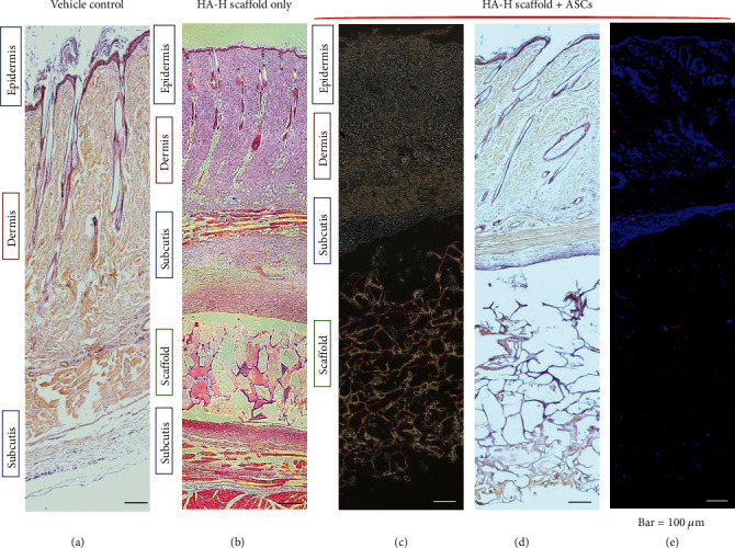
Figure 5.

Histology images of wound beds. (a) Normal rat skin with paraffin section; (b) HA-H matrix was implanted into the rat skin. (c–e) ASC HA-H porous matrix was implanted within the rat skin. (c) Viewed with white light, (d) histochemical staining with the paraffin section, and (e) histochemical staining with the frozen section in fluorescent light. ASCs were dyed in PKH26 with a red color, and the blue color was a matrix surrounding tissue cells with DAPI staining.
