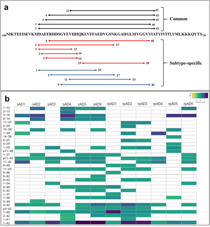
Fig. 2.
Diversity in proteoforms identified in formic acid-soluble fraction of sAD and rpAD. Top-down MALDI-MS identified 33 different proteoforms of Aβ. Although intersubject variability is evident in proteoform signature obtained from various cases, Aβp3-42, Aβ3-42 Aβp11-42, Aβ11-42, Aβ4-42, Aβ1-40, and Aβ1-42 were the most dominant proteoforms. (a) The sequence of proteoforms common in sAD (red), rpAD (blue), or both groups (black) is marked on APP (only sequence between amino acid 660 to 730 is shown). (b) The heatmap depicts the relative intensities of all identified proteoforms, calculated using the average area under the curve (AUC) from five measurements. The intensities were normalized for each sample and the respective Z-scores of proteoforms were used for this plot

